Polyclonal Antibodies The original way to produce antibodies injecting an animal with the antigen and collecting the serum is a cheap and easy way to generate multiple antibodies to a given target. In this case the researchers chose 05 micro liters.
Learn About Immunostaining With Antibodies To Characterize Cells And Tissues Cell Signaling Technology
Check the antibodys datasheet on the manufacturers website for available pictures of IF stainings and compare them with.
. Localization of patient antibodies is visualized by a second fluorescein antihuman IgG antibody evaluated under a fluorescence microscope. 2 Dermal pattern consistent with epidermolysis bullosa acquisita. For example if the primary antibody is raised in mouse the second antibody should be select as goat-anti-mouse antibody if the primary antibody is raised in rabbit the second antibody should be select as goat-anti-rabbit antibody.
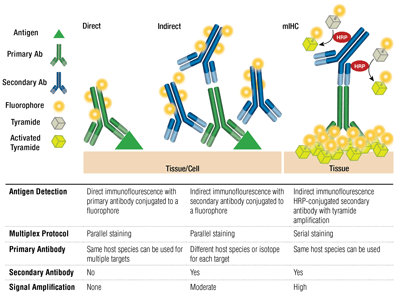
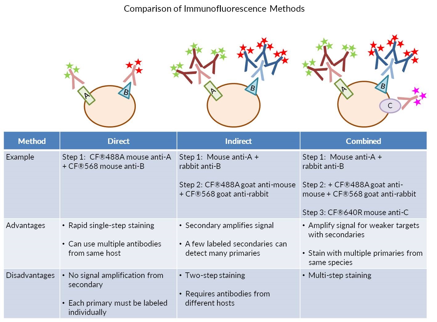
It is most important that select second antibody according to the source of the primary antibody. We use cookies to enhance your experience. Direct IF indirect IF and combined IF.
It is most important that select second antibody according to the source of the primary antibody. If infection with a parasite is suspected and blood film stool or urine examinations are either not indicated or are negative then the appropriate serology test for specific IgG. Ad Without Leaving Home Check Yourself or Child for Protective Antibodies to Prior Infection.
You can add the solution and incubate in the same way as the primary. Formalin-fixed Paraffin Embedded FFPE Slide De-paraffinization and rehydration. Secondary antibodies can be engineered to carry different-colored fluorophores ie.
2The fluorescent marker on second. Identify whether you want an antibody that will recognize the whole protein a certain segment of it eg N- or C-termini or a specific peptide sequence within. Controls are often the most.
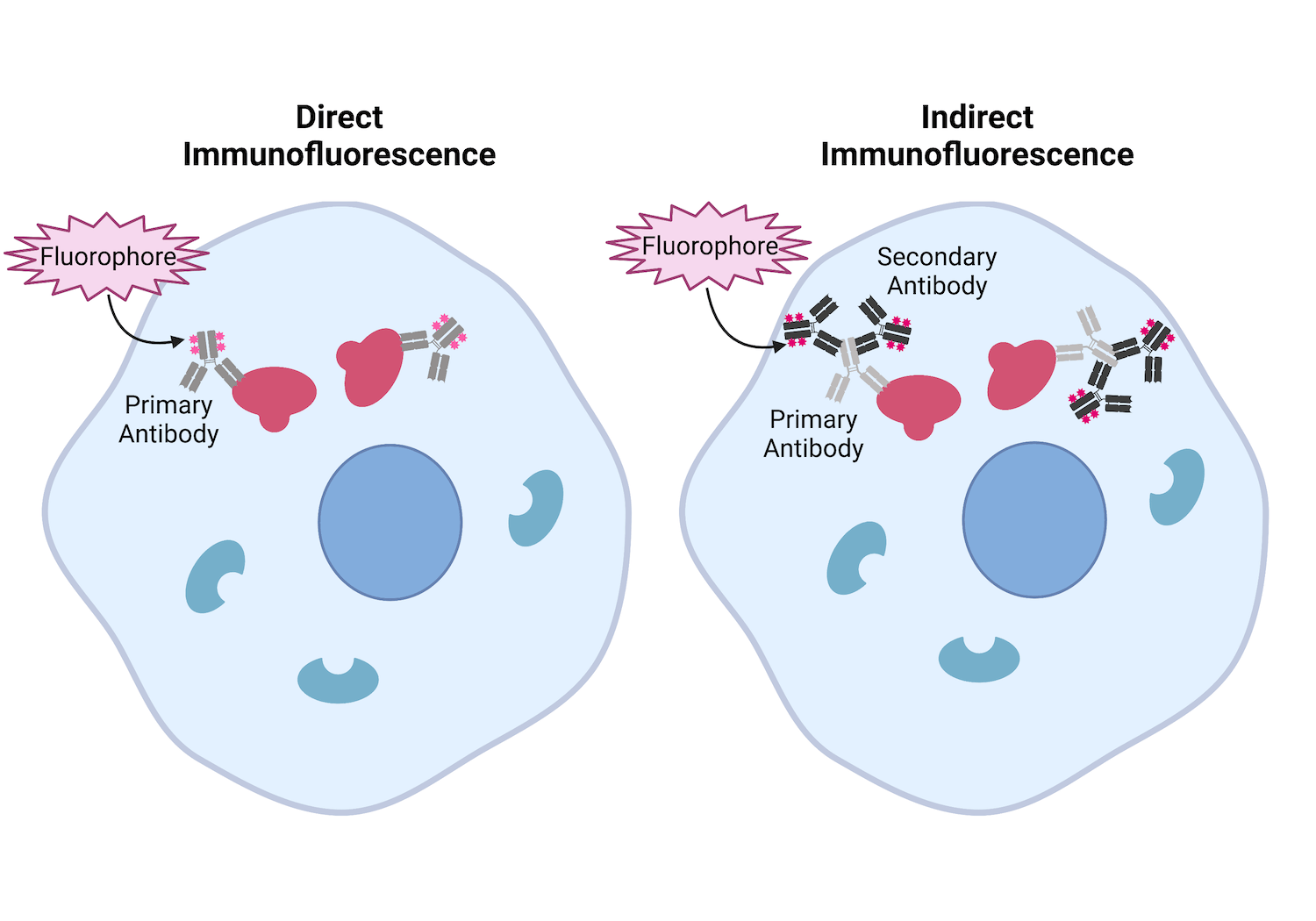
Secondary indirect immunofluorescence uses two antibodies. Primary antibody that specifically binds to epitope and a matched secondary antibody conjugated with fluorescence dye. Direct primary or indirect secondary.
Most secondary antibodies can be used after 1200-1500 dilution for 1 hour at room temperature or overnight at 4 C. The unlabeled first primary antibody specifically binds the target molecule and the secondary antibody which carries the fluorophore recognizes the primary antibody and binds to it. Here we describe preparation of specimens preserved in different types of media and step-by-step methods for both direct and indirect immunofluorescence staining.
If serum contains basement zone BMZ antibodies on split-skin substrate patterns will be reported as. Thus high concentrations of these protein competitors can out-compete your antibodies and lower your background noise. Any type of immunofluorescence IF technique will require the use of a fluorescent-labeled antibody that is used to identify and located proteins antigens or other biological molecules present.
Pipetting errors a little less or a little more will not change the SI. The idea is that your antibodies will not bind to your nonspecific epitopes any better than these nonspecific protein competitors will. How to choose second antibody for IF in immunofluorescence IFICC.
There are disadvantages of this method especially in the case of using polyclonal reagents in flow cytometry. Dilute the fluorophore-conjugated secondary antibody in PBS-005 Tween 20 again you can include 5 goat serum if the secondary antibodies are from goat. Staining index versus antibody concentration.
This procedure was found to be more sensitive than the immunofluorescence procedure and in addition it was much easier to determine when a. Test Kit Checks if You or Your Child Had SARS-Co-V-2 Now Have Protective Antibodies. Report includes presence and titer of circulating antibodies.
Dual Labeling Using Fluorescence. 1 Epidermal pattern consistent with pemphigoid. Multiple secondary antibodies can bind a single primary antibody.
Place slides in 60C oven for 30 min or overnight at 37C. Use positive controls different cells that contain protein Use several different antibodies that are directed against same protein Preabsorption controls. Indirect IF is using two antibodies for the staining.
See Note 3 Incubate sections in two 100 mL. Prepare controls containing only 01 saponinPBSFBS or if available containing preimmune antiserum if rabbit polyclonal antibody is being used or specific primary antibody with the antigen added in excess. Choose your antibody according to already used and validated primary antibodies in literature on the subject.
For antibody A the maximal separation is somewhere between 025 and 1 micro liters. Negative in normal individuals. Sino Biological gives a detailed solution to help you troubleshoot this problem.
The use of two antibodies to visualize an antigen provides the advantage of picking what color will represent the location of the antigen. Immunofluorescence assays are antigen detection assays that generally require use of a fluorescent microscope to produce results in approximately 2-4 hours with moderate sensitivity and high specificity. Mixing Ab with protein or antigenic peptide before labeling to eliminate binding.
The detection of specific IgM and IgA antibodies may be of value in determining the approximate time of initial infection with Toxoplasma gondii but is not recommended for any other parasitic disease. Incubate 24 μm thick sections in three 100 mL washes of xylene for 5 minutes each for a total of 15 minutes. At this concentration if there are any issues in the preparation ie.
Islet cell antibodies have been detected using the procedure of indirect immunofluorescence. Both direct DFA and indirect fluorescent antibody IFA staining assays are available to detect influenza A and B viral antigens in. There are two methods available depending on the scope of the experiment or the specific antibodies in use.
We have adopted an immunohistochemical procedure using glucose oxidase to the histochemical identification of islet cell antibody. Direct IF is using a single primary antibody that is conjugated with fluorescent dye. Also preincubating cells with antigenic peptide Use antigen-negative cells cells from knock-out mouse.
For example if the primary antibody is raised in mouse the second antibody should be select as goat-anti-mouse antibody if the primary antibody is raised in rabbit the second antibody should be select as goat-anti-rabbit antibody. Choose your antibody according to already used and validated primary antibodies in literature on the subject. 2The fluorescent marker on second.
However at 025 micro liters if there is an issue and. Chinese中文简体 Chinese中文繁體 Japanese日本語 Korean한국어 215-583-7898 Leave a Message. Fluorescence antibody FA immunofluorescence staining is a reliable and powerful technique for a wide range of research and diagnostic purposes.
There are three types of IF. This method is often the most economical and often works well for monoclonal antibodies. Check the antibodys datasheet on the manufacturers website for available pictures of IF stainings and compare them with your expectations or other published illustrations.

Our Comprehensive Guide To Performing Double Immunofluorescence

Direct Vs Indirect Immunofluorescence Abcam
Considerations For Immunofluorescence Staining Biotium
Antibodies 101 Introduction To Immunofluorescence


0 Comments